PET Skanning makes use of some quantities of radio-substances known as radio-tracers or -pharmaceuticals, a peculiar digicam and a pc to consider organs functionings. PET detects modifications occurring at cellular range, which helps to identify the disease at the stage of its inception – much earlier than is possible with other methods.
PET-CT Method
PET-CT – complex technique combining PET and CT. Implementing such technique, you can get detailed data about development of malignant neoplasms.
- With CT, specialists take several x-rays of the body, then the system combines them into one, creating a 3D model.
- PET uses a drug with a low degree of radioactivity to identify those cells showing abnormal behavior.
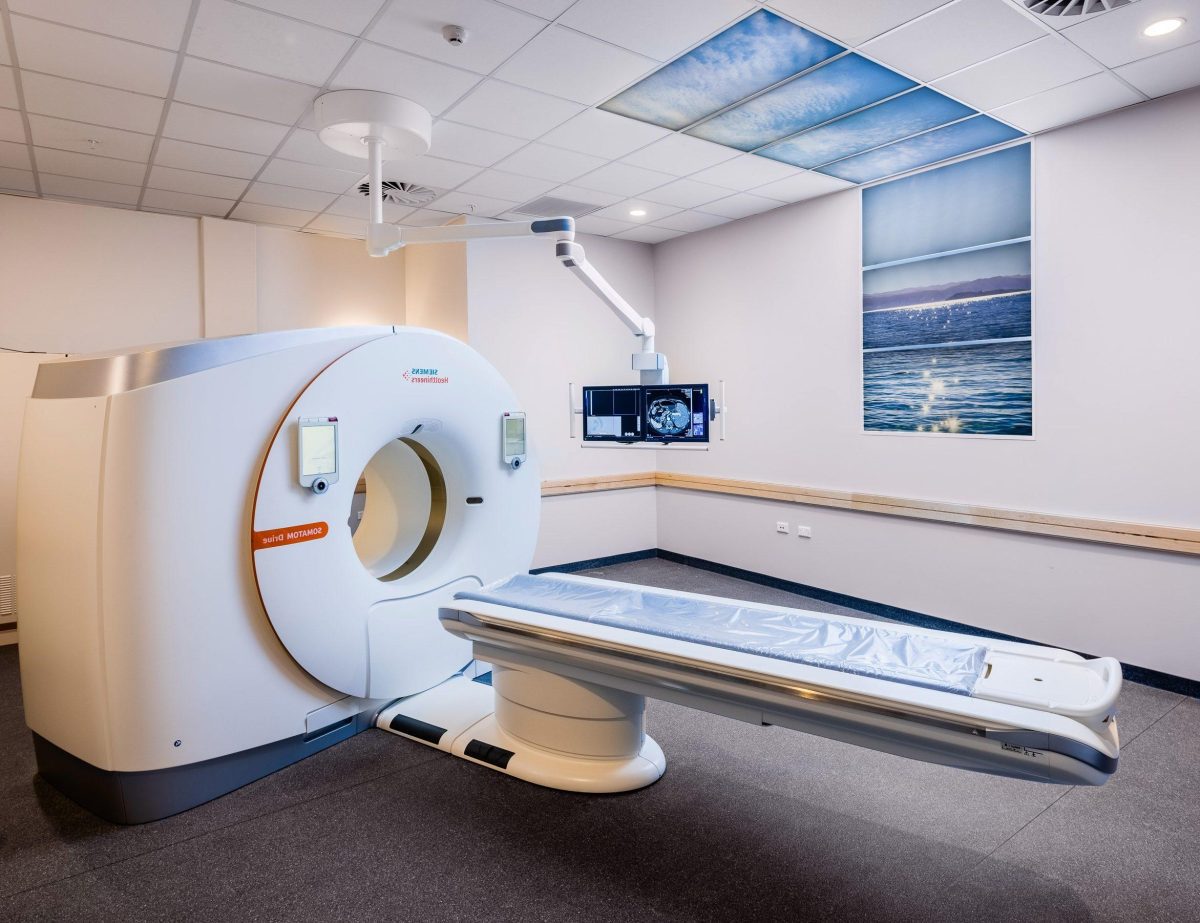
PET-CT scan, for the most part, is performed outpatiently in the radiology department. Radiologists work with special scanners – usually, process lasts from half an hour to 60 min.
Positron emission tomography and computed tomography purposes
These methodologies apply in next mentioned cases.
- Detection of the presence of cancerogenic formations, and affected zone determination.
- Getting accurate diagnostic outcomes.
- Evaluation of the effectiveness of the chosen therapy.
- Determining the likelihood of relapse.
- Assessment of metabolic processes and viability of body systems.
- Determination of how the patient’s heart attack affected areas of the heart muscle.
- Identification of areas of the heart that can be affected by angioplasty or where coronary bypass surgical manipulations can be performed.
- Detection of abnormality present in cerebrum.
- Creating a map of heart and cerebrum operational abilities.
What Can Experts See with PET-CT & PET?
- Evaluation of options vital to the organism: bloodflow, sugar indicators and oxygen consumption frequency.
- Determining which systems are not operating properly.
- Detection of cancer cells, which helps prevent metastasis.
- Evaluation of the effectiveness of the therapy program – if necessary, therapy is adjusted.
- Detection of cancerogenic pathologies.
- Assessment of the size of the tumor and the area of its spread.
- Obtaining data, based on its possible drawing guessing about need for surgical manipulations.
- Choice of method of treatment.
- Assessment of the risk of recurrence.
- Planning of radiotherapy.
Using PET-CT, it is possible to reveal effectiveness of therapy aimed at combating carcinoma. Technology demonstrates areas where cancerogenic cells still remain.
CT VS PET Scan
CT uses x-rays, MRI uses radio and magnetic waves. Both techniques enable specialists to create a holistic still image of body structures. PET uses an active radio substance to demonstrate operable ability of organs and the behavior of cells in real time. Thereby, difference between PET and CT scan is first mentioned helps to reveal pathological modifications in organism earlier.
PET operational function has below described action mechanism:
Prior to PET-CT scanning, you’ll get injection of radiosugar substance in small quantity referred to as fluorodeoxyglucose-18. Most frequent radiotracer is F-18 fluorodeoxyglucose (FDG) – molecule comparable to glucose. This substance is occasionally referred to as FDG-18, radio-glucose, or tracer. Cancerogenic cellules are extra metabolically lively and can also soak up glucose at a greater level. Greater active reaction can be viewed on PET outcomes. It gives your specialists possibility to discover ailment earlier than it can be detected on different visualizing tests.
Preparing for PET-CT
As for preparing basics, going to PET-CT scan, it’s necessary to limit meal getting within 6 hrs before. You’re permitted to drink water. Sometimes doctors also recommend refraining from physical exercises.
For diabetic patients who can’t stay without food, it’s necessary that you adjust your nutrition – no-food hours fall into a certain time before. Specialists must consult patients.
Methodology Implementation
It’s as follows:
- Patients get on the desk. If it’s needed, catheter will be inserted in patient’s vein – on the forearm or arm.
- Radio substance – FDG-18 – injection is made.
- As a rule, for the disclosure of its properties, it’s needed 60 min to drug. Patients are at rest, silently.
- Sometimes, people are given a bit of contrast agent localized in the intestine – this gives radiologists the possibility to better interpret diagnosis outcomes.
- The person is then transferred to a PET-CT machine.
Mostly, process time is no more than 60 minutes.
Patients Feelings
Mostly, this process is acheless, with exception of intravenous injection. Sometimes, patients report mild discomfort.
Pros & Negatives
Begin with positive aspects.
- PET-CT scan allows specialists to obtain unique data about the patient’s anatomy and the functions of organ systems.
- Nuclear methodologies provide medical professionals with most detailed data about diagnosis or therapy of various benign and malignant ailments.
- Cheaper and super accurate method compared with exploratory surgery.
- Abnormal modifications determination at cellular level – pathology becomes definable at a very early stage.
- High detailing.
- Low error probability.
Now let’s look at some of the risks.
- A small radiative substance influences organism – radio agent is introduced during process implementation.
- Allergic responses are possible, however, they manifest themselves slightly.
- Contraindications to the use of this process: pregnancy and lactation.
- After it, slight redness and itching may appear on skin.