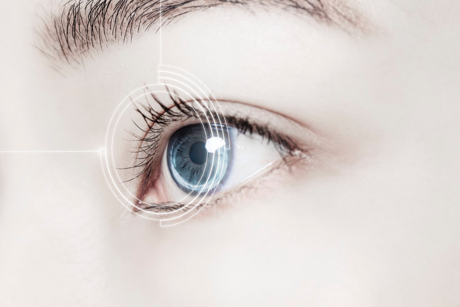
In astigmatism, anterior transparent part of the eyeball is not completely round in shape. The human eyeball, in its ideal natural state, has the shape of an absolutely round ball. However, if the shape of the cornea is somehow disturbed, the light falling on it begins to refract more in one direction or another. Only part of the object is in focus, therefore, objects being at some length can take on wavy or blurry outlines.
In many cases, astigmatism is paired with farsightedness or nearsightedness. It can be corrected with glasses, lenses, or surgically.
Causes of astigmatism
What is astigmatism? When cornea are not perfectly round, resembling an egg with two mismatched curves, then the light that hits them is not refracted equally. Thus, different images are formed on their surface, overlapping each other, or merging with each other. This leads to blurry and blurred vision. It is in the case when the lenses have a greater contortion in one of directions that astigmatism develops.
This defect can be in people from birth, or it can arise as a result of trauma or surgery. Contrary to popular belief, astigmatism is not caused and cannot be aggravated by reading in low light or sitting too close to the TV.
Symptoms of astigmatism
General astigmatism symptoms are as follows:
- distortion or blurring;
- rapid eye fatigue, especially when focusing on an object for a long time;
- severe headaches due to tension in the eyes;
- deterioration of vision and perception of the picture at night.
Diagnosis of astigmatism
The symptomatology of this defect manifests itself slowly: a person begins to gradually notice certain changes in vision. To identify the possible presence of the disease, a complete eye examination is carried out, in particular, a visual acuity test. In addition, doctors can resort to the following diagnostic techniques.
- Phoropter – a patient viewing several lenses in order to determine which of them give the most accurate picture.
- Keratometer – measurement of the curvature of the cornea central part and how well it can focus on objects.
- Autorefractor – directing lights into the eye and measuring the degree of its change when reflected from the back wall. This allows specialists to choose the right lenses.
- Corneal topographer – obtaining detailed information about cornea shape and complete data on its general condition.
Treatment of astigmatism
Special lenses cope with most cases of astigmatism. However, if in addition to this defect, which is observed in a mild form, patients no longer have vision problems, glasses and lenses may not be necessary.
In the usual form of this defect, lenses, glasses are required, or, in some cases, minor surgical correction can be resorted to. Doctors recommend starting to wear glasses and lenses gradually, helping your eyes get used to the new state. With severe defects, rigid lenses are prescribed, which are also worn during sleep. They correct the shape of the cornea.
Sometimes, doctors resort to laser surgery, which changes the shape of the cornea – PRK or LASIK. Another surgical technique used for this defect is astigmatic keratotomy, or limbal incisions, where tiny incisions are made in the sharp curves to focus light more accurately.


